Used Dental Digital Panoramic X-Ray | i-CAT Classic
Current Listings
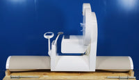


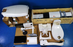
The i-CAT Classic® (i-CAT Imaging System) is a Cone Beam Computed Tomographic and Panoramic imaging system. It is designed for straightforward installation, ease of maintenance and service. It is a high frequency, constant potential, fixed anode X-Ray source. It incorporates a flat panel, amorphous silicon image detector at 20 cm x 25 cm, with a cesium iodide conversion layer
The i-CAT® is connected to a desktop computer to provide the user interface and data processing.
Features:
- 3D volumetric images
- True anatomic measurements
- 14 BIT gray scale
- Machine is the size of a traditional panoramic X-Ray
- Fast 20 second scan time
- High resolution for all views
Benefits:
- Surgical predictability
- Save time w/ 1 scan vs. multiple traditional X-Ray
- Digital for paperless office
- Can be used in multiple dental applications
- Increase patient confidence and boost sales by increased case acceptance
- Keep business in-house vs. outside specialist referral
Limitations/Considerations:
- Higher default radiation dose than newer models
- Because of fewer field of view options
- Does not automatically adjust volume sizes for children
- Does not provide iPAN Ceph 2D, but this can be captured from 3D. (low radiation dose panoramic came in Next Generation Model)
Technical Specifications:
-
Power: 120 or 240V Ac
-
Hertz: 60-50Hz
-
Tube Voltage: 120 kVp
-
Tube Current: 3-7 mA
-
Line Voltage Regulation requirement: + 10%
-
Line Current: 10 Amps (115V) or 5 Amps (230V)
-
Weight: 426 lbs
-
Keyboard Dimensions: (43 cm Length) x (18 cm Width)
-
Monitor with Stand Dimensions: (45 cm Height) x (34 cm length)
-
Tower Computer Dimensions: (43 cm Height) x (47 cm Length) x (22 cm Width)
-
Field of View: 16 cm(Diameter) 13-22 cm (Height)
-
Source to Sensor distance: :27 inches (68.58 cm)
-
Source to Patient distance: 18 inches (45.72 cm) (center of rotation)
-
Voltage Wave shape: Constant Potential
-
Scan Time: 40/20/10 Seconds
-
X-Ray Source: 120 kVp 3-7 Ma (pulsed)
-
Software: Xoran Cat, iCAT Vision & 3DVR, TxStudio powered by Anatomage
-
Focal Spot: 0.050038 cm (0.0197 inches)
-
Maximum Deviation:kVp: + 5 kVp
RELATED EQUIPMENT