Used Dental Digital Panoramic X-Ray | i-CAT Next Generation 3D ( 1043 )
Current Listings
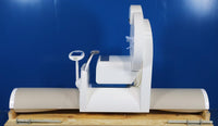


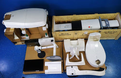
The Cone Beam Volumetric Tomography and Panoramic X-ray device is used for dental head and neck applications. The system consists of a Scanner and Computer Workstation which is suitable for an in-office environment. This scanning device is an open design that allows Patients to sit upright during a procedure. An electric powered seat is built into the device for proper Patient positioning.
The system consists of a Scanner and Computer Workstation. In order for the system to operate, both the Scanner and Computer Workstation must be turned ON. The system captures data for 3D Skull Reconstruction for the following procedures:
- Implants
- TM Joints
- Reconstructed Panoramic
- Reconstructed Cephalometrics
- Airway / Sinus, etc. • Nerve Canal
- PAN - Optional Conventional Digital Panoramic Feature
Cone Beam Volumetric Tomography is a medical imaging technique that uses X-rays to obtain cross-sectional images of the head or neck. Quality of the images depends on the level and amount of X-ray energy delivered to the tissue. Imaging displays both highdensity tissue, such as bone, and soft tissue. When interpreted by a trained Physician, these images provide useful diagnostic information.
- CONE BEAM 3D
- With i-CAT Next Generation
- Dynamic clinical control
- LARGE 3D FOV
- With 14 x 8 cm field of view
- The Extended Field of View, 16cm (d) x 22cm (h), captures full cranial height images.
- Standard Scan: 4, 6, 8, 10, 13 cm (h) x 16 cm (d), 8 cm (h) x 8 cm (d) Extended Field of View (Cephalometric): 17 cm (d) x 23 cm (h) Custom Mode: any height from 2-13 cm (h) x 16 cm (d).
- Adjustable 3d FOV
- 2-in-1 functionality
- Fully compatible with SureSmile software with a super-fast 14.9 second scan time
- TX studio software
- Delivers clean, clear high quality 3D scans and panoramic images
- Provides views of soft tissue and crisp visualization of hard tissue and bone structures
- Allows to acquire high-definition 3D scans while minimizing patient dose
- Fast scan protocol
- Convenient benefit for practices that require both 2D and 3D cone beam imaging
- Provides all the tools dentists and dental specialists need to diagnose and plan implants, restorations, surgical procedures, endodontics, orthodontics, TMJ and airway assessment
- Laser beams can cause optical damage. Instruct the Patient to close eyes to avoid looking into the beam. The use of optical instruments such as eyeglasses with large diopter or mirrors, increase eye hazard with this product.
- The useful and scattered beams can produce serious or fatal bodily injuries to Patients and persons in the surrounding area if used by an unskilled operator. Adequate precautions must always be taken to avoid or reduce exposure to the useful beam or to scattered radiation.
-
Footprint: 48 x 70 x 36 in
-
Scan Time: 5 s, 9 s, 27 s
-
Patient Positioning: Seated, Wheelchair Accessible
-
Primary Reconstruction Time: Less than 30 s
-
Volume Reconstruction: Yes
-
Panoramic Reconstruction: Yes
-
Cephalo Reconstruction: Yes
-
DICOM Export: DICOM Export
-
ACTUAL DIMENSIONS: 48.13"(W) x 52.58"(D) x 69.51"(H)
-
Image Detector: Amorphous Silicon Flat Panel Sensor with Csl Scintillator
-
Gray Scale: 14 bit
-
Voxel Size (Slice Thickness): 0.125 to 0.4 mm
-
POWER REQUIREMENTS: 110V/15A
-
Total Weight: 510 lbs. (231.3 kg)
RELATED EQUIPMENT